
By Peggy Bush
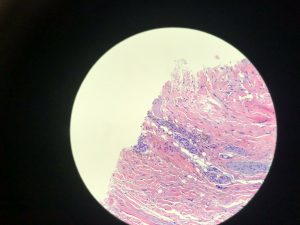
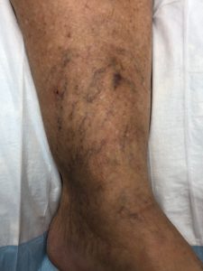
There are two types of staining that can occur when treating telangiectasia. Type I is diffuse and results from vessel wall lysis after sclerotherapy. There is red cell extravasation to the reticular dermis and iron pigment remains after red cell lysis.
Type II staining occurs with intra-luminal thrombosis. It is distinguished by a linear stain in former vessel lumen and is very common when foam sclerotherapy is used. Evacuation of thrombus early may be helpful, but some staining will persist.
In most patients, staining will subside in 3-6 months. Patients with skin types 4-6 may take up to a year to resolve and this may be due to increased melanocyte production as well as iron deposition secondary to an inflammatory response.
Staining can be treated with heat modalities, although this can be somewhat limited in effectiveness. Lopez, et al, 2001, concluded that weekly subcutaneous administration of DM 500 mg reduces the time to depigmentation by 82 percent in patients with post-sclerotherapy hyperpigmentation. This is rarely needed but can be considered in patients with severe persistent staining.
Ronald Bush, MD, FACS, has found with histological examination that patients with staining may have in the treated vessel or in the reticular dermis hemosiderin deposition. This is usually cleared by macrophages and there may also be leukocytes in the area, which is indicative of an inflammatory response. Staining is iron pigment that occurs after red cell lysis.

TIPS TO AVOID STAINING
Use the lowest concentration of sclerosant when injecting veins and this includes foam sclerotherapy. Empty the spider vein with manual compression after injection and prevent refilling if possible. Flushing with bacteriostatic water may also be helpful. Consider using subdermal tumescent to reduce the lumen size during the post treatment period.
Cosmetically, staining is unacceptable to patients. Plant-based creams may be helpful, especially products that contain licorice root extract oil. Licorice root extract is nature’s most potent lightener. The extract from the licorice root is naturally high in glabridin and this chemical inhibits tyrosinase.
Other complications and treatment will be discussed at the March 18 Aesthetic Vein Course at Venous Symposium in New York City. Complete information on the 2020 Venous Symposium is available at venous-symposium.com/ . VTN
![]() Peggy Bush is an advanced practice registered nurse working with Dr. Ronald Bush in clinical treatment and research at Water’s Edge Dermatology, a practice with 36 locations throughout Florida.
Peggy Bush is an advanced practice registered nurse working with Dr. Ronald Bush in clinical treatment and research at Water’s Edge Dermatology, a practice with 36 locations throughout Florida.
REFERENCES
Lopez L, Dilley RB, & Henriquez, JA. Cutaneous hyperpigmentation following venous sclerotherapy treated with deferoxamine mesylate 2001;(9):795-8.
Photography Courtesy of Water’s Edge Dermatology, 2019
